STRATIS
The aim of this Challenge is to produce a human-relevant and high throughput in vitro or ex vivo platform that recapitulates the complex structures of skeletal muscle and the pathology of significant injury to them. The platform must offer a model for novel wound therapeutics and approaches to restore form and function after significant soft tissue injury.
Challenges briefing webinar
View the Challenges briefing webinar recording to find out more about this Challenge.
Phase 2 awarded
A team led by Professor Mark Lewis, Loughborough University, has been awarded £1 million to deliver the project 3D assays of vascularised and innervated human skeletal muscle injury for functional screening of pro-regenerative therapeutics.
Phase 1 awarded
Three Phase 1 Awards were made to project teams led by:
- Dr James Dixon, University of Nottingham, £99,088.
- Professor Mark Lewis, Loughborough University, £91,590.
- Dr Yung-Yao Lin, Queen Mary University of London, £100,000.
Challenge Launched
Sponsored by Dstl and co-finded by Dstl and EPSRC this Challenge aims to produce a human-relevant and high throughput in vitro or ex vivo platform that recapitulates the complex structures of skeletal muscle and the pathology of significant injury to them. The platform must offer a model for novel wound therapeutics and approaches to restore form and function after significant soft tissue injury.
Background
Volumetric muscle loss (VML) is the substantive loss of skeletal muscle and impairment of regenerative capability that can occur as a result of trauma, surgery or degenerative disease. Wounds of this nature are complex, involving skin, muscle and associated structures such as microvasculature, nerves and the extracellular matrix. In addition, volumetric wounds caused by trauma – particularly blast and ballistic injuries – can evolve, resulting in loss of tissue beyond that lost as a result of the primary insult. VML wounds often exhibit sub-optimal healing, with a poor response to reconstructive efforts and resulting in long-term scarring. Current strategies for reconstruction of VML wounds focus on preservation of the tissue that remains, with a return of full function often unachievable. Loss of form and function, particularly in the limbs, manifests as severe permanent deficits that adversely impact on physical and mental rehabilitation.
A variety of clinical regenerative and tissue engineering strategies designed to replace soft tissue are under investigation, including novel materials, either seeded with cells or coated with materials that stimulate endogenous tissue growth (O'Brien, 2011; Zhu et al., 2019). Physiological and functional integration of these constructs is challenging, and it is likely that therapeutic interventions to stimulate the survival and proliferation of existing structures alongside tissue reconstruction will be required. Due to the complex interplay between the structures within soft tissue during healing and regeneration, preclinical models to evaluate reconstructive and regenerative approaches for VML need to include muscle, vasculature and where possible, skin.
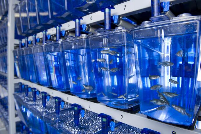
In vivo models
In vivo VML models have a full complement of cell types and tissue architecture and allow long-term studies which is critical when looking at regenerative responses. However, there are differences in animal and human pathophysiology associated with wound healing, particularly in rodents, which makes translation to humans difficult (Ansell et al., 2012; Grada et al., 2018). For example, mice have an additional muscle layer that is mostly absent in humans – this enables their skin to move independently of the deeper tissues and allows wound healing via contraction rather than the re-epithelialisation that is seen in humans. In vivo models are usually not suitable for high throughput screening of therapeutic candidates, and the severity and duration of experiments which involve creating surgical wounds create ethical concerns.
Ex vivo models
Ex vivo approaches including explant and perfused systems using animal and human tissue are being explored for both skin (Mendoza-Garcia et al., 2015; Ternullo et al., 2017) and to a lesser extent muscle (Park et al., 2012; Smith and Meyer 2019). These approaches are advantageous in terms of inclusion of relevant cell types and tissue architecture and are amenable to wounding and the testing of novel therapeutics (Mendoza-Garcia et al., 2015). However, they are limited in the length of time they can be kept physiologically viable.
In vitro models
Commonly-used 2D in vitro platforms only model simple wounds such as the scratch test (Cory, 2011) and therefore do not recapitulate the complexity of VML or translate well to the clinic (Ansell et al., 2012; Grada et al., 2018). These models are cheap, high throughput and can include primary human and animal skin or muscle cells but usually only employ a limited number of cell types, lack tissue architecture and do not support functional or mechanical readouts or manipulations (Cory, 2011).
Complex 3D in vitro models of skin and muscle have been developed that are amenable to long-term culture, contain multiple cell types and tissue architectures, and retain the ability to support higher throughput studies and functional readouts (Afshar et al., 2020; Matei et al., 2019). The availability of models however, that contain innervated muscle and skin and are applicable to the study of wounding, wound healing and regeneration is limited. The Horizon 2020 MyoChip project aims to develop an innervated and vascularised muscle-on-a-chip model but it is unclear what functional outcomes will be achieved and whether it will be suitable for wounding and other mechanobiological investigations.
This Challenge aims to build on the emerging capabilities in the area of complex 3D cultures and regenerative medicine to develop a human cell or tissue-derived 3D in vitro or ex vivo model of vascularised skeletal muscle, with additional tissue architecture, for example, skin and/or innervation that replicates the mechanical properties of native tissue and is suitable for ‘wounding’ and the study of subsequent regenerative processes.
3Rs benefits
Models of muscle loss are invasive and often involve surgically exposing the muscle of interest (e.g. tibialis anterior or biceps femoris) and creating a defect using a biopsy punch before closing the wound and allowing the animals to recover from the anaesthesia. These models cause pain and distress, even when analgesics are used, and the welfare of the animal is closely monitored. A typical study for testing engineered tissues and supportive materials and biological therapeutics uses five experimental groups (six to eight rats per group) and a control group (16 rats).
The development of a more complex in vitro or ex vivo model of wounding to muscle could replace a significant number of animal models in the screening and testing of regenerative therapeutics by providing a higher throughput option for tissue engineering strategies using, for example, small molecules or gene therapy candidates. This could improve the early evaluation of approaches and, optimise translation.
The market for advanced wound care products is extensive and expected to grow from $10.8 billion USD in 2019 to $15.56 billion USD by 2027 (Fortune Business Insights, 2020). Within this market, the development of a complex in vitro or ex vivo model of wounding and wound healing could also be adapted for other acute wounding modalities, for example, burns, ischaemia or chronic wounds, using appropriate tissue or primary cells. This has the potential to replace animals typically used in such studies that involve simple incisional/excisional wounds to more complex crush injuries, involving electroporation, myotoxin administration or ischaemia.
Full Challenge information
Assessment information
Review Panel membership
|
Institution |
|
|
University of Sunderland |
|
| Dr Abigail Spear (Sponsor) | Dstl |
|
University of Leeds |
|
|
VU University Medical Center |
|
|
Hull York Medical School |
|
|
Imperial College NHS Trust |
|
|
University of Birmingham |
|
|
Independent |
|
|
University of Bradford |