NC3Rs | 20 Years: Pioneering Better Science
CRACK IT Challenge

EASE: Design, Fabrication and Testing of a Mouse Embryo Culture Chip
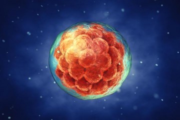
An early stage embryo

At a glance

Completed
Award date
January 2017 - June 2019
Contract amount
£ 95,883
Sponsor(s)

R

  • Refinement

Contents

Overview

Genetically altered (GA) mice are used extensively to study the function and regulation of genes and their role in human development and disease. The generation of GA mouse models involves one of three techniques that use embryos at different stages of development: (i) pronuclear injections; (ii) in vitro fertilisation (IVF); or (iii) embryonic stem cell injection. Embryos from all three approaches are transferred into pseudopregnant recipients primarily using surgical techniques performed under general anaesthesia.

In recent years, Non-Surgical Embryo Transfer (NSET) techniques have been developed for trans-cervical transfer of blastocysts, morulae, DNA-microinjected embryos, and embryonic stem cell-containing embryos in the uterine cavity. NSET technology avoids surgery and improves animal welfare. However, the NSET techniques can only be used to transfer late stage pre-implantation embryos, and current approaches for extended culture of one and two-cell embryos generated from IVF programmes or following pronuclear injection severely compromises their implantation success. This means that these common techniques do not get used in conjunction with the current NSET systems. Further optimization of murine embryo culture techniques is required to enable the use of NSET approaches with all stages of embryo to improve implantation and pregnancy rates.

The team at University of Leeds led by Dr Virginia Pensabene proposes to develop a novel and reliable microfluidic Organ-On-a-Chip (OoC)device to significantly improve the developmental competence of in vitro-derived mouse embryos and so maximise implantation rates and increase the efficiency of NSET. Using OoC devices will allow Dr Pensabene to control specific features of cell culture (cell position, flow, mechanical cues), and provide unprecedented flexibility in dissecting the cellular, molecular, chemical and physical factors which contribute to the development and function of the embryos.

This project will couple expertise in microfluidic, microfabrication technology and reproductive biology (Dr Pensabene) with the extensive experience of Professor Helen Picton in IVF and embryology to create a “bioinspired” OoC. These microfluidic devices will reduce embryo exposure to in vitro stressors and improve the health and competence of manipulated embryos through to the blastocyst stage through the better manipulation of media volume and composition, pH and oxygen tension.

Full details about this CRACK IT Challenge can be found on the CRACK IT website.