InPulse
The aim of this Challenge was to generate a physiologically-relevant contractility platform with cells that are phenotypically ‘mature’, possess a robust contractile apparatus, move calcium between intracellular and extracellular spaces and metabolically generate substantive amounts of energy.
To address the Challenge the team led by Professor Chris Denning, University of Nottingham, have developed a suite of assays to assess cardiac liabilities of drugs using human induced pluripotent stem cell-derived cardiomyocytes (hiPSC-CMs).
10 years of CRACK IT webinar: Putting in vitro platforms at the heart of cardiotoxicity testing
In July 2021, we held a webinar highlighting the technologies and platforms developed to address the InPulse CRACK IT Challenge through developing a human-based in vitro system for cardiotoxicity testing. Speakers include Professor Chris Denning (University of Nottingham), who led the team that delivered the InPulse Challenge, and Dr Peter Clements (GSK), lead Sponsor of the Challenge. The recording of the webinar is now available online, as part of our 10 years of CRACK IT celebrations.
Publication
Hall C et al. (2021). Complex Relationship Between Cardiac Fibroblasts and Cardiomyocytes in Health and Disease. Journal of the American Heart Association 10: e019338. doi: 10.1161/JAHA.120.019338
Publication
Bhagwan JR et al. (2020). Isogenic models of hypertrophic cardiomyopathy unveil differential phenotypes and mechanism-driven therapeutics. Journal of Molecular and Cellular Cardiology 145: 43–53. doi: 10.1016/j.yjmcc.2020.06.003
Challenge completed
Through the InPulse Challenge, the team led by Professor Chris Denning, University of Nottingham, have developed a suite of assays to assess cardiac liabilities of drugs using human induced pluripotent stem-cell derived cardiomyocytes (hiPSC-CMs).
Conference presentation
Safety Pharmacology Society 2018 Annual Meeting (Washington, USA)
Simultaneous Measurement of Contraction, Voltage and Calcium in hiPSC-CMs for the Detection of Inotropic Effects under Blinded Conditions.
Publication
Berend J. van Meer, Luca Sala, et al. (2018). Quantification of Muscle Contraction In Vitro and In Vivo Using MUSCLEMOTION Software: From Stem Cell-Derived Cardiomyocytes to Zebrafish and Human Hearts. Current Protocols in Human Genetics 99(1): 67. doi: 10.1002/cphg.67.
Publication
Mosqueira D, Mannhardt I, Bhagwan JR, et al. (2018). CRISPR/Cas9 editing in human pluripotent stem cell-cardiomyocytes highlights arrhythmias, hypocontractility, and energy depletion as potential therapeutic targets for hypertrophic cardiomyopathy. Eur Heart J 39(43): 3879–3892. doi: 10.1093/eurheartj/ehy249.
Publication
Ulmer BM, Stoehr A, Schulze ML, et al. (2018). Contractile Work Contributes to Maturation of Energy Metabolism in hiPSC-Derived Cardiomyocytes. Stem Cell Reports 13;10(3): 834-847. doi: 10.1016/j.stemcr.2018.01.039.
Publication
Smith JG, Owen T, Bhagwan JR, et al. (2018). Isogenic Pairs of hiPSC-CMs with Hypertrophic Cardiomyopathy/LVNC-Associated ACTC1 E99K Mutation Unveil Differential Functional Deficits. Stem cell reports 11(5): 1226-1243. doi.org/10.1016/j.stemcr.2018.10.006.
Publication
Sala L, van Meer BJ, Tertoolen LGJ, et al. (2018). MUSCLEMOTION: A Versatile Open Software Tool to Quantify Cardiomyocyte and Cardiac Muscle Contraction In Vitro and In Vivo. Circ Res 2;122(3): e5-e16. doi: 10.1161/CIRCRESAHA.117.312067.
Products launched
Physiologically-relevant cardiomyocyte models and associated technologies developed through the Challenge are now available.
Publication
Duncan G, Firth K, George V, et al. (2017). Drug-Mediated Shortening of Action Potentials in LQTS2 Human Induced Pluripotent Stem Cell-Derived Cardiomyocytes. Stem Cells Dev 1;26(23): 1695-1705. doi: 10.1089/scd.2017.0172.
Conference presentation
Safety Pharmacology Society Annual Meeting 2017 (Berlin, Germany)
Studying Inotropic Compound Effects in Human iPSC-derived Cardiomyocytes using 2D and 3D Models.
Conference presentation
Safety Pharmacology Society Annual Meeting 2016 (Vanouver, Canada)
Poster 1: Development of an in vitro platform using physiologically mature human induced pluripotent stem cell-derived cardiomyoyctes to screen for drug-induced effects on cardiac contractility.
Poster 2: Characterization of electrical and mechanical functions of Pluricyte® hiPSC derived cardiomyocytes using the optical platform CellOPTIQ.
Publication
van Meer BJ, Tertoolen LG, Mummery CL (2016). Measuring Physiological Responses of Human Pluripotent Stem Cell Derived Cardiomyocytes to Drugs and Disease. Stem Cells 34(8): 2008-15. doi: 10.1002/stem.2403.
Publication
van Meer BJ, de Vries H, Firth KSA, et al. (2016). Small molecule absorption by PDMS in the context of drug response bioassays. Biochem Biophys Res Commun 8;482(2): 323-328. doi: 10.1016/j.bbrc.2016.11.062.
Publication
Rajamohan D, Kalra S, Duc Hoang M, et al. (2016). Automated Electrophysiological and Pharmacological Evaluation of Human Pluripotent Stem Cell-Derived Cardiomyocytes. Stem Cells Dev 15;25(6): 439-52. doi: 10.1089/scd.2015.0253.
Publication
Eder A, Vollert I, Hansen A, et al. (2016). Human engineered heart tissue as a model system for drug testing. Adv Drug Deliv Rev. 15(96): 214-24. doi: 10.1016/j.addr.2015.05.010.
Publication
Kosmidis G, Bellin M, Ribeiro MC, et al. (2015). Altered calcium handling and increased contraction force in human embryonic stem cell derived cardiomyocytes following short term dexamethasone exposure. Biochemical and biophysical research communications 467(4): 998-1005. doi: 10.1016/j.bbrc.2015.10.026.
Publication
Birket MJ, Ribeiro MC, Kosmidis G, et al. (2015). Contractile Defect Caused by Mutation in MYBPC3 Revealed under Conditions Optimized for Human PSCCardiomyocyte Function. Cell Rep. 13(4) :733-745. doi: 10.1016/j.celrep.2015.09.025.
Publication
Denning C, Borgdorff V, Crutchley J, et al. (2015). Cardiomyocytes from human pluripotent stem cells: From laboratory curiosity to industrial biomedical platform. Biochim Biophys Acta 1863: 1728-48. doi: 10.1016/j.bbamcr.2015.10.014.
Publication
Birket MJ, Ribeiro MC, Verkerk AO, et al. (2015). Expansion and patterning of cardiovascular progenitors derived from human pluripotent stem cells. Nat Biotechnol 33(9): 970-9. doi: 10.1038/nbt.3271.
Publication
Patel AK, Celiz AD, Rajamohan D, et al. (2015). A defined synthetic substrate for serum-free culture of human stem cell derived cardiomyocytes with improved functional maturity identified using combinatorial materials microarrays. Biomaterials 61: 257-65. doi: 10.1016/j.biomaterials.2015.05.019.
Publication
Ribeiro MC, Tertoolen LG, Guadix JA, et al. (2015). Functional maturation of human pluripotent stem cell derived cardiomyocytes in vitro--correlation between contraction force and electrophysiology. Biomaterials 51: 138-150. doi: 10.1016/j.biomaterials.2015.01.067.
Phase 2 awarded
A team led by Professor Chris Denning, University of Nottingham, has been awarded £999,915 to deliver the project: Engineered 2D & 3D hiPSC-CM platforms to detect cardiovascular safety liabilities.
Phase 1 awarded
Three Phase 1 Awards were made to project teams led by:
- Professor Chris Denning, University of Nottingham, £100,000.
- Professor Cesare Terraciano, Imperial College London, £99,958.
- Professor Wolfram Zimmerman, University Medical Center Göttingen, £100,000.
Challenge launched
Sponsored by GSK, the InPulse Challenge aims to generate a physiologically-relevant contractility platform with cells that are phenotypically ‘mature’, possess a robust contractile apparatus, move calcium between intracellular and extracellular spaces and metabolically generate substantive amounts of energy.
Background
Cardiovascular (CV) safety liabilities are one of the most common causes of drug attrition in both the nonclinical and clinical setting. These liabilities manifest in a variety of ways that include structural injuries and derangements in CV function. Changes in contractility may lead to clinical heart failure and even death. Important classes of drugs, such as anti-neoplastic tyrosine kinase inhibitors, are currently facing particular safety challenges with this effect.
iPS cell-derived cardiomyocytes (iPSC-CMs) have shown early promise for replacing animal use in cardiovascular research and many pharmaceutical companies and academic researchers are developing assay platforms to identify potential electrophysiological and structural liabilities earlier in development (i.e. ‘pre-animal’). A similar capability for contractility assessments would be very useful from a scientific and 3Rs perspective.
Historically, there has been a heavy reliance on highly invasive, technically demanding, surgically instrumented telemetered animal models in preclinical studies that provide indirect measures of contractility in vivo. More recently, imaging (e.g. echocardiography) has been used as an investigative and mechanistic tool both preclinically and clinically but it is low throughput and labour intensive. The only in vitro/ex vivo models that are available are perfused whole heart models from animals and isolated cardiac cells from animals and humans. There is no currently validated/qualified human-derived in vitro system for assessing drug-induced changes in contractility under varying levels of physiologic load.
The ability to study changes in cardiac contractility in vitro would allow the investigation of potential causes of long QT syndrome that are both hERG and non-hERG dependent. This would allow a more accurate prediction of potential drug-induced cardiac toxicity in preclinical and clinical settings; thus help to reduce attrition. Mechanistic understanding of changes in cardiac contractility from in vitro models would also be relevant to efficacy studies earlier in the drug discovery process.
The rapid development of human iPS cell technologies and materials sciences provides an opportunity to develop a dynamic, human-relevant assay system to screen and characterize novel drug candidates for cardiac contractility liabilities. Recent publications have demonstrated that the extracellular matrix, morphology and orientation of cardiac cells are important for cardiac contractility and calcium signalling in cardiac myocytes and there have been significant advances in substrates for monitoring contractile tension.However, there is the need for a robust in vitro model that reflects the 3D architecture of cardiac tissue with mature cell phenotypes.
3Rs benefits
- Assessment of developmental drug pre-candidates on cardiac contractility relies exclusively on the use of animals prior to clinical trials. A typical study uses 12 to 24 dogs.
- A physiologically relevant 3D in vitro model will replace the use of animals in these studies and reduce the number of animals needed to assess the mechanism of action of any drug-induced effect.
- An in vitro system where load can be adjusted to model the pathophysiology of disease tissue could also be used to reduce animal use in efficacy studies. A typical efficacy study uses approximately 24 mice.
- Once validated, an in vitro platform could be used to screen compounds and provide earlier go/no-go decisions hence reducing the number of animals used to test compounds that would ultimately fail in development.
Phase 1 winners
Project teams led by:
- Professor Chris Denning, University of Nottingham, £100,000.
- Professor Cesare Terraciano, Imperial College London, £99,958.
- Professor Wolfram Zimmerman, University Medical Center Göttingen, £100,000.
Phase 2 winner
Project team led by:
Full Challenge information
Through the InPulse Challenge, the team have developed a suite of assays to assess cardiac liabilities of drugs using hiPSC cardiac myocytes (CMs).
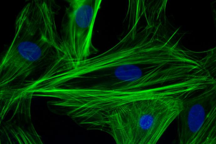
The team compared different lines of hiPSC-CMs, including PluriCyte cells from Ncardia, in combination with different platform technologies, each tested through the same set of compounds provided by the Sponsor to establish the optimum configuration for assessment of the effects of drugs on cardiac toxicity with a focus on contractility.
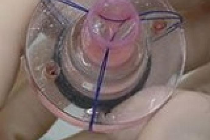
Two hiPSC-CMs configurations were selected to provide a comprehensive final assay set: a 2D monolayer cell platform to provide high throughput readouts and a 3D engineered heart tissue system (EHT Technologies) that is lower throughput but can replicate better the cardiac microenvironment to produce higher levels of cell maturity and provide more detailed mechanistic insights (Figure 1).
Using the 2D monolayers of hiPSC-CMs, two technology platforms were utilised to provide assay read outs:
- The Triple Transient Measurement System (TTM) developed by Leiden University that can continually measure contractility, Ca2+ handling and electrophysiology within a repeating 3 msec window (Figure 2).
- The CellOPTIQ system developed by Clyde Biosciences in Glasgow that can measure contractility, Ca2+ handling and electrophysiology from the same region of the well.
Assessment of the effects of drugs on the contraction of hiPSC-CMs EHTs is performed via calculation of absolute force from the extent of deflection of two silicon posts, whilst calcium handling is reported using a lentiviral genetically encoded calcium indicator.
The team also developed MUSCLEMOTION- a versatile open software tool to quantify cardiomyocyte and cardiac muscle contraction in vitro and in vivo. This single software platform can be used to quantify contractility in:
- Single adult rabbit cardiomyocytes.
- hiPSC-CM as single cells, monolayers, cardio spheres and EHTs with or without drug or disease challenge.
- In vivo healthy or diseased zebrafish hearts.
- Human MRI scans from patients who were healthy or with deficiency in heart function.
The InPulse Challenge has led to greater validation of the hiPSC-CM model in cardiovascular safety testing and produced a wealth of knowledge and data to inform the optimal choice of cardiac cell platform to assess contractility.
For further information please contact Professor Chris Denning in the first instance.
Assays to assess the cardiac liabilities of drugs
Multiple different technologies and platforms have been developed or advanced as products of the InPulse Challenge. These include three-dimensional engineered heart tissues (EHT Technologies) and fully functional human induced pluripotent stem cell-derived cardiomyocytes for preclinical safety assessment (NCardia) and a new software platform for the measurement of cardiomyocyte and cardiac muscle contraction (MuscleMotion).
EHT Technologies
Engineered heart tissues (EHTs) are three-dimensional, hydrogel-based muscle constructs that can be generated from isolated heart cells of chicken, rat, mouse, human embryonic stem cells (hESC) and human induced pluripotent stem cells (hiPSCs) to replace in vivo studies.
The new generation of fibrin-based miniaturised EHTs uses fibrin (fibrinogen/ thrombin) as a hydrogel, standard 24-well culture dishes, rectangular casting molds in agarose, and two elastic silicone (PDMS) posts per well to which the growing muscle tissue attaches.
Muscle contractions (either spontaneous or electrically stimulated) deflect the posts and can be evaluated by video-optical recording.
EHTs will start coherent beating after approximately five-seven days and can be used for:
- Drug screening.
- Disease modeling.
- Basic cardiovascular research.
You can find out more about this product on the Dinabios website.
MUSCLEMOTION: a versatile open software tool to quantify cardiomyocyte and cardiac muscle contraction in vitro and in vivo
There are several methods to measure cardiomyocyte and muscle contraction, but these require customized hardware, expensive apparatus, and advanced informatics or can only be used in single experimental models. Consequently, data and techniques have been difficult to reproduce across models and laboratories, analysis is time consuming, and only specialist researchers can quantify data.
Musclemotion is a versatile open software tool to quantify cardiomyocyte and cardiac muscle contraction in vitro and in vivo. This single software platform can be used to quantify contractility in:
- Single adult rabbit cardiomyocytes.
- hiPSC-CM as single cells, monolayers, cardio spheres and EHTs with or without drug or disease challenge.
- In vivo healthy or diseased zebrafish hearts.
- Human echocardiograpic recordings.
You can find out more about this product on the Musclemotion website.
NCardia
Preclinical studies of drug effects with human induced pluripotent stem cell (hiPSC)-derived cardiomyocytes provide novel possibilities for the assessment of efficient and reliable safety and efficacy drug screenings and avoiding the use of animals. Pluricyte® Cardiomyocytes are fully functional hiPSC-derived cardiomyocytes obtained without any genetically modification or purification/selection procedures.
Pluricyte® Cardiomyocytes are cultured in Pluricyte® Cardiomyocyte Medium, a serum free, well-defined culture and maturation medium.