NC3Rs | 20 Years: Pioneering Better Science
Fellowship

Multiphoton imaging in human liver tissues: validation of a new tool for drug discovery
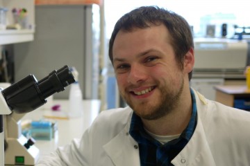
Dr Scott Davies

At a glance

Completed
Award date
September 2018 - December 2020
Grant amount
£109,410
Principal investigator
Dr Scott Davies
Institute
University of Birmingham

R

  • Reduction
  • Replacement
Read the abstract
View the grant profile on GtR

Overview

Chronic liver disease affects over 300 million people worldwide and transplantation is often the only available cure. Mouse models of liver disease are moderate/severe and often fail to yield clinically translational data. This is, in part, due to the variances in immune cell function between mouse and human. Imaging mice via intravital microscopy has allowed for real-time in vivo insights into liver immune cell function. However, these are severe procedures and still present difficulty with clinical application. There is a pressing need for translational models of liver disease.  

During my PhD, I studied how hepatocytes engulf dead cells. This process (efferocytosis) caused hepatocytes to fail cell division in vitro and become multinucleate. I also observed that in mice with ischemia reperfusion injury, hepatocytes next to necrotic legions had more nuclei. To prove that efferocytosis caused this, we set up a model whereby donor human livers were perfused with an inhibitor of efferocytosis and then injured, which prevented multinucleation of hepatocytes near the injury. This proved that perfused human tissue could be used to replicate murine in vivo observations. Our group has also shown that regulatory T cells (Treg) were captured alive by and deleted by hepatocytes. As Tregs have shown potential for dampening liver disease, halting their deletion by hepatocytes may prove of benefit. 

This proposal aims to improve the real-time imaging of liver immune cells while adhering to the 3Rs of animal testing. We aim to refine the imaging of mT/mG mouse livers by comparing in vivo intravital microscopy to ex vivo multiphoton imaging of livers for Treg-hepatocyte interactions in the presence of inhibitor C1. We shall then establish the first use of multiphoton microscopy for imaging perfused human liver and testing drug efficacy. This model will encourage others to test compounds on human liver disease tissue, yielding more clinically relevant results.

Impacts

Publications

  1. Davies SP et al. (2020). Cell-in-Cell Structures in the Liver: A Tale of Four E’s. Frontiers in Immunology 11:650. doi: 10.3389/fimmu.2020.00650