Rapid adoption of self-sustaining 3D epithelioid models: Replacing mice in epithelial tissue and cancer studies
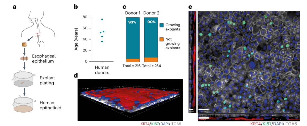
Dr David Fernandez-Antoran’s team at the University of Cambridge have transferred an epithelioid model to other cancer research groups with joint CRUK-NC3Rs funding. David’s method exploits the wound healing properties of epithelia and allows long-term, self-maintaining and expandable primary 3D cultures of mouse and human epithelial tissues from different origins. The long-term culture of primary epithelial cells has historically been challenging. Organoids using cells sourced from animal or human tissues exist, however, they continuously expand, and therefore do not attain the balance between proliferation and differentiation that maintains adult tissues in a steady state.

David’s model, which he developed during his tenure as a Junior Group Leader in CRUK’s Radiation Research Network is a significant innovation. The epithelioid cultures faithfully replicate in vivo tissue organisation and gene expression and self-maintain for at least a year. They are also amenable to gene editing technologies and treatment with chemotherapy and radiotherapy. David’s lab has replaced its use of mice by generating 3D epithelioids from human tissues (replacing the use of 200 mice per year).
The CRUK-NC3Rs award has allowed the transfer of the epithelioid model to four other labs in Cambridge and its qualification for various epithelial tissue types including the oesophagus, trachea, skin, ovary and bladder – with all of the labs either fully replacing or reducing their animal use as a result, saving approximately 400 mice per year. The funding has also allowed the model to be adopted by groups in Spain and Malaysia with similar reductions in animal use and the biotechnology company, Omnipotent Biosciences Limited, have adopted the epithelioid model for studying ageing and tissue regeneration. The epithelioid model has been published in Nature Genetics and David, together with colleagues in RadNet, has received almost £300k of further funding from Cancer Research UK, and an additional £100k of grants from other sources. Building on the development of the epithelioid model, David’s team are now able to culture both normal and tumour tissues, enabling them to assess the differential responses of these tissues to radiotherapy treatments in order to design and optimise cancer therapies. They have obtained funding to study the response and evolution of head and neck tumours under radiotherapy treatments, using patient-derived epithelioid cultures, in collaboration with NHS UK and the universities of Manchester and Oxford.
"David’s epithelioid model has huge potential to replace animal use across multiple cancer types, including of the oesophagus, trachea, skin, nasopharynx, ovary, bladder, lymph nodes and head and neck."
Dr Katie Bates, Head of Funding

