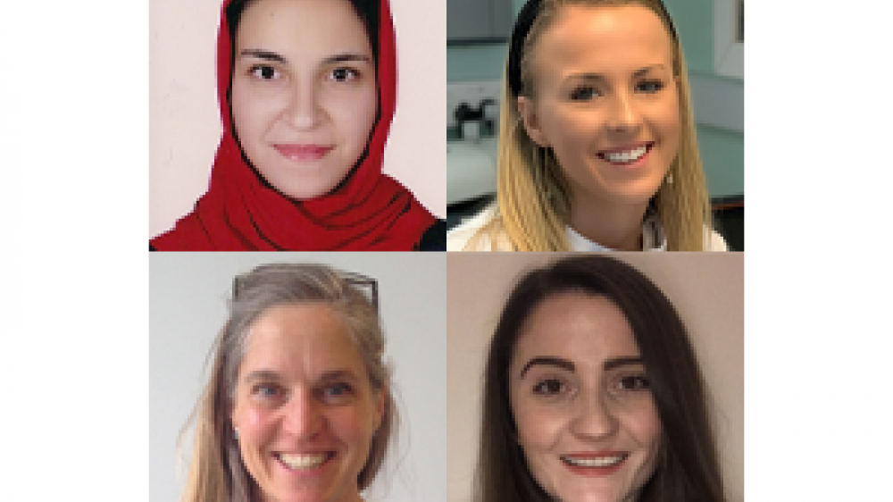
Meet our newest Training Fellows

We have awarded four new NC3Rs Training Fellowships, totalling £512k, to support the development of talented 3Rs-minded postdoctoral researchers.
The Training Fellows will focus on replacing, refining and reducing animals in research whilst addressing key scientific questions, encompassing computational models of extracellular matrices, a microfluidic in vitro model of the retina, the effects of X-rays in laboratory animals and a zebrafish model of congenital myopathy.
The Training Fellowship scheme was launched in 2016 to provide early career researchers with an opportunity to acquire new skills and research experience focused on the 3Rs, with the support and guidance of a dedicated mentor.
More information on our latest Training Fellowship awardees can be found on our Meet our Training Fellows page.
The latest awards are summarised below:
Dr Nargess Khalilgharibi, University College London – A computational framework to investigate the mechanical role of the extracellular matrix in tissue development and disease
Organoids can reduce reliance on animal models, but most current organoid culture techniques use extracellular matrix (ECM) derived from animals. The composition of animal-derived ECM is highly variable, which affects experimental reproducibility and translation of results, so synthetic alternatives are being developed. Nargess will establish an open-source, multiscale computational platform that can model organoid growth in different synthetic ECM, making it easier for researchers to choose the optimum ECM for their work.
Dr Savannah Lynn, University of Southampton – MacuSIM: A microfluidic, in vitro model of the outer retina as an experimental platform for macular disease and therapeutic trials
Age-related Macular Degeneration (AMD) is a common eye disease that results in the gradual loss of central vision due to damage to the macula (the most sensitive area in the centre of the retina). Different species are used as models to investigate the underlying causes of AMD and develop new drugs. This includes rodents, which are commonly used despite not having a macula. Savannah is developing a microfluidic in vitro 3D model of the retina, mimicking the architecture of the human macula and modelling blood flow. She will use this model to investigate AMD disease pathology and potential therapeutics.
Dr Wendy McDougald, University of Edinburgh – Modelling radiobiology effects of X-rays in small laboratory animals to develop guidelines for preclinical computed tomography imaging
In the clinical setting, human exposure to X-rays is tightly controlled and regulated. This is not the case for preclinical research, meaning animals are frequently overexposed to X-rays, particularly in longitudinal imaging studies. Overexposure to radiation can cause cellular damage, affecting welfare and confounding experimental findings. Using computational modelling and a 3D-printed rodent model, Wendy will work to develop preclinical guidelines to refine radiation exposure protocols whilst maintaining high-quality images.
Dr Rhiannon Morgan, University of Liverpool – A novel embryonic zebrafish model to replace mammals in the study of 3-hydroxyacyl CoA dehydratase 1-associated muscle disorders
Rhiannon’s project will focus on generating a zebrafish model of congenital myopathy, which affects humans and dogs and leads to muscle wasting and weakness. Congenital myopathy, caused by mutation in the HACD1 gene, is poorly understood and currently untreatable. Zebrafish are a well-established non-mammalian model for studying muscle development and disease. Rhiannon will use non-protected stage zebrafish embryos to investigate how muscle structure and function are altered by HACD1 deficiency, replacing and reducing the use of dogs and mice.
