£100k awarded in new CRACK IT Solutions funding
Two innovative projects have been awarded £50k each through the NC3Rs CRACK IT Solutions technology partnering programme.
CRACK IT Solutions supports collaborations to help further develop and validate novel technologies with potential 3Rs benefits.
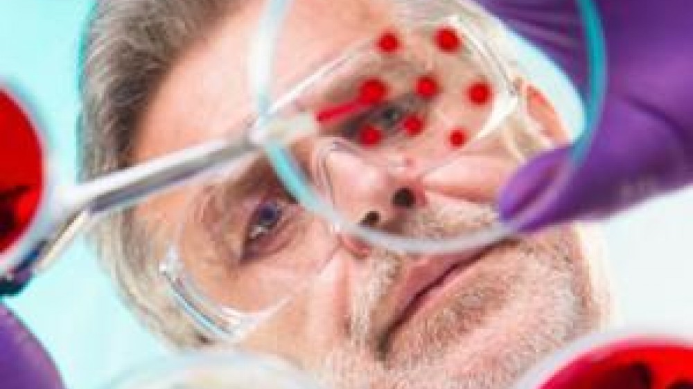
Applying novel imaging platforms to tissue engineered corneal tissue constructs
Professor Zhihong Huang and colleagues at the University of Dundee have developed functional optical coherence tomography (OCT) to enable high quality, ultrafast, qualitative and quantitative characterisation of both tissue structure and function in vivo. The technique has the potential to both reduce and refine animal use, as long-term non-invasive serial tissue monitoring in small animals is possible. There is also the possibility to use OCT on biomimetic tissue models, enabling more structural and functional information to be generated in these models without destroying the tissue, potentially opening up new opportunities for these non-animal models to replace some in vivo work.
Through CRACK IT Solutions, Professor Huang engaged a number of potential new collaborators. She is now working with Thea Pharmaceuticals and Keele University on a CRACK IT Solutions-funded project to explore the potential use of OCT in monitoring corneal wound healing in engineered corneal tissue constructs.
Multiple animal models for corneal wound healing are currently used including rodent, rabbit and dog models. These can be painful due to the high density of nerves in the eye and can result in infections. Thea Pharmaceuticals has recently developed a new drug to promote corneal ulcer healing and they will use OCT to monitor the effect of this drug on tissue repair and regeneration in the non-animal corneal tissue model.
Studying pain ex vivo
Professor Adalberto Merighi and his team from the Department of Veterinary Sciences, University of Turin, Italy has developed a cultured spinal cord slice model that can provide unique insight into the central mechanisms of pain.
Through CRACK IT Solutions, Professor Merighi sought collaborators to help further develop and validate these spinal cord slice platforms (SCSPs) for studying pain ex vivo, and for preclinical development of novel therapeutics. With CRACK IT Solutions funding, he is now working with a team led by Professor Eustace Johnson at the University of Chester, UK. Together, they will use the SCSP model to study the effects of transplanting mesenchymal stem/stromal cells (MSCs), which have been proposed as a clinical treatment for spinal cord injury, into the spinal cord to better understand their effects on neuroinflammation, central neuropathic pain and neuronal hyperexcitability.
Traditionally these experiments would have been carried out in vivo in large numbers of mice, causing substantial suffering to the animals involved. In one published example, 453 adult mice were used to show that early transplantation of MSCs after spinal cord injury relieves pain hypersensitivity. However, using this system instead has the potential to reduce the number of animals used by up to 50%, as multiple slices can be cultured from a single animal.