NC3Rs | 20 Years: Pioneering Better Science
PhD Studentship

Mapping mitochondrial contact sites during neuronal ageing and neurodegeneration in Drosophila
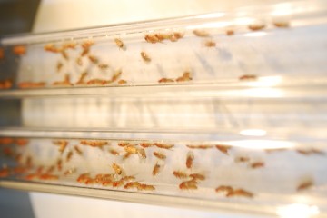
Drosophila melanogaster (fruit flies) in two clear plastic tubes.

At a glance

In progress
Award date
February 2023 - January 2027
Grant amount
£120,000
Principal investigator
Dr Alessio Vagnoni

Co-investigator(s)

Institute
King's College London

R

  • Replacement

Overview

Why did we fund this project?

This award aims to further develop a fruit fly (Drosophila) model to investigate the role of mitochondria in neuronal ageing and neurodegenerative disease and replace some experiments in mice and zebrafish.

The contacts made between mitochondria and other subcellular organelles are important for neurons to function. Specifically, the interaction between mitochondria and the endoplasmic reticulum (ER) is important for ageing and neurodegeneration. Currently these contacts are visualised using electron microscopy of dissected mice and zebrafish tissues. In a previous NC3Rs-funded project, Dr Alessio Vagnoni developed an alternative approach, using green fluorescent protein to visualise the contacts between mitochondria and the ER in the nervous system of live Drosophila. Current scientific thinking suggests that Drosophila do not experience suffering and so can act as a partial replacement of vertebrates. The student, with Dr Alessio Vagnoni, will build confidence in the Drosophila model and demonstrate its utility in studying ageing and neurodegenerative diseases by investigating whether mitochondrial interactions with other organelles (peroxisomes and lysosomes) contribute to abnormal neuronal function.

Application abstract

The interactions that the mitochondria establish with other cellular compartments is central to neuronal functioning and much focus has been devoted to the mechanisms regulating mitochondria-endoplasmic reticulum (ER) contact sites and their role in ageing and neurodegeneration. Supported by an NC3Rs SKT grant, we had previously produced a genetically encoded split-GFP probe to study mitochondrial-ER contacts during ageing, which has proved to be important for the field and created several opportunities for collaboration. Beyond the ER, however, mitochondrial establish contacts with several other subcellular organelles. The interactions with the peroxisomes and the lysosomes constitute a particularly important, although still poorly explored, cellular functional hub that has only recently begun to be studied in detail. Whether these interactions are perturbed in the context of ageing and neurodegeneration is not known. To study this aspect, we now intend to go one step further by developing and validating genetically encoded probes for the analysis of the associations that the mitochondria establish with the lysosomes (Mito-Lyso) and the peroxisomes (Mito-Pex). We will do so by combining these new sensors, inspired by the split-GFP technology, with super-resolution microscopy to measure Mito- Lyso and Mito-Pex contacts during ageing and in Drosophila models of Alzheimer’s disease, Parkinson’s disease and amyotrophic lateral sclerosis.

We recently discovered that reduced mitochondrial motility is a fundamental regulator of neuronal homeostasis during ageing in Drosophila, although the underlying mechanisms are still poorly understood and an important question in the field. An exciting possibility is that reduced mitochondrial transport may play a role in contact formations during ageing and in neurodegeneration. We will test this hypothesis by coupling our powerful genetic tools to our advanced methodology for imaging of live neurons in the intact nervous system of adult Drosophila.

We have established a wide network of collaborators who work with mice and zebrafish and study the cell biology of the neurons and mitochondrial contacts in different contexts. We will maximise the uptake of our technology by leveraging our existing network and the structure that we previously put in place to successfully transfer our Drosophila platform to collaborators. A direct and tangible output of this project will be able to replace approximately 120 mice and 200 adult zebrafish within one year of project completion by transferring our technology to international collaborators. In a concurrent and related study, a local collaboration has resulted in the reduction of approximately 160 mice otherwise used for breeding and tissue dissection, and there is the possibility to also transfer the tools developed through this studentship to local researchers, which would augment the potential for local reduction. Overall, the project will advance our basic understanding of the cell biology of neuronal ageing with a clear path to reduce the number of vertebrates used for this type of studies.

Finally, there is a considerable appetite for a technology that would facilitate the study of mitochondrial contacts during ageing in a physiological context. The gold standard to study mitochondrial contact sites in animal tissues is electron microscopy (EM), which is time-consuming and requires a specialised and expensive set-up and cannot be used for live imaging. Therefore,  providing easy-to-use, versatile genetically encoded tools would reduce the reliance on EM studies and further increase the transfer-ability of our system and the potential for replacement of vertebrate models.