NC3Rs | 20 Years: Pioneering Better Science
Skills and Knowledge Transfer grant

Transfer of the epicardial-cardiac organotypic culture model to support the ex vivo screening of gene therapy candidates
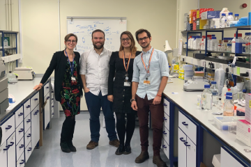
Dr Paola Campagnolo on the left with three colleagues on the right side of her in a laboratory

At a glance

Completed
Award date
August 2019 - March 2022
Grant amount
£75,562
Principal investigator
Dr Paola Campagnolo
Institute
University of Surrey

R

  • Replacement
Read the abstract
View the grant profile on GtR

Impact story

This project has been featured in an impact story, showcasing how NC3Rs-funded research comes together to develop innovative new 3Rs tools and technologies that are pioneering better science.

Overview

Why did we fund this project?

This award aims to replace rodents in gene therapy studies for cardiac regeneration with the EpCardio-TS model, an ex vivo porcine cardiac slice model.

Investigating gene therapy strategies in the epicardium, the most external layer of the heart, typically uses mice or rats. The genes of interest are over-expressed, and a myocardial infarction induced under general anaesthetic, to determine the effects of the gene on cardiac regeneration. Inducing a myocardial infarction is classified as severe under the UK’s Animals (Scientific Procedures) Act 1986 due to the level of suffering caused. EpCardio-TS was developed by Dr Paola Campagnolo with NC3Rs funding and uses tissue obtained either as a by-product from the abattoir or as surplus material from animals killed in the laboratory for another scientific purpose. The slices are viable in culture, metabolically active and provide a 3D model with multiple relevant cell types that is a better representation of the human heart than other cellular in vitro methods, which are often 2D and include only one cell type. Paola has used the model in her own laboratory to study epicardial cell proliferation, migration and differentiation.

Using nanoneedles, Paola has demonstrated the localised transfer of reporter genes to fluorescently label the epicardial cells and by optimising a decolouration protocol has been able to visualise the labelled epicardial cells within the slice, including during cell migration. Professor Molly Stevens at Imperial College London and Dr Ofelia Martinez-Estrada from the University of Barcelona will collaborate with Paola to facilitate the adoption of EpCardio-TS into their gene therapy studies.

Publications

  1. Matos R et al. (2023). Ex Vivo Perfusion Culture of Large Blood Vessels in a 3D Printed Bioreactor. J. Vis. Exp. (197):e65465. doi: 10.3791/65465
  2. Maselli D et al. (2022). Porcine Organotypic Epicardial Slice Protocol: A Tool for the Study of Epicardium in Cardiovascular Research. Front. Cardiovasc. Med. 9. doi: 10.3389/fcvm.2022.920013
  3. Maselli D et al. (2022). Epicardial slices: an innovative 3D organotypic model to study epicardial cell physiology and activation. NPJ Regen Med 7:7. doi: 10.1038/s41536-021-00202-7
  4. Matos R et al. (2022). 3D Printed Bioreactor Enabling the Pulsatile Culture of Native and Angioplastied Large Arteries. Front. Cardiovasc. Med. 9. doi: 10.3389/fcvm.2022.864580
  5. Cheung Y et al. (2020). A Critical Role for Perivascular Cells in Amplifying Vascular Leakage Induced by Dengue Virus Nonstructural Protein 1. mSphere 5. doi: 10.1128/msphere.00258-20
  6. Mastrullo V et al. (2020). Angiogenesis in Tissue Engineering: As Nature Intended? Front. Bioeng. Biotechnol. 8. doi: 10.3389/fbioe.2020.00188