Using clinical inspiration to refine small animal radiotherapy
Dr Karl Butterworth and PhD Student Dr Kathryn Brown have applied high precision cancer radiotherapy techniques used to treat human patients to improve the reproducibility and translatability of preclinical animal trials.
From patients to preclinical studies
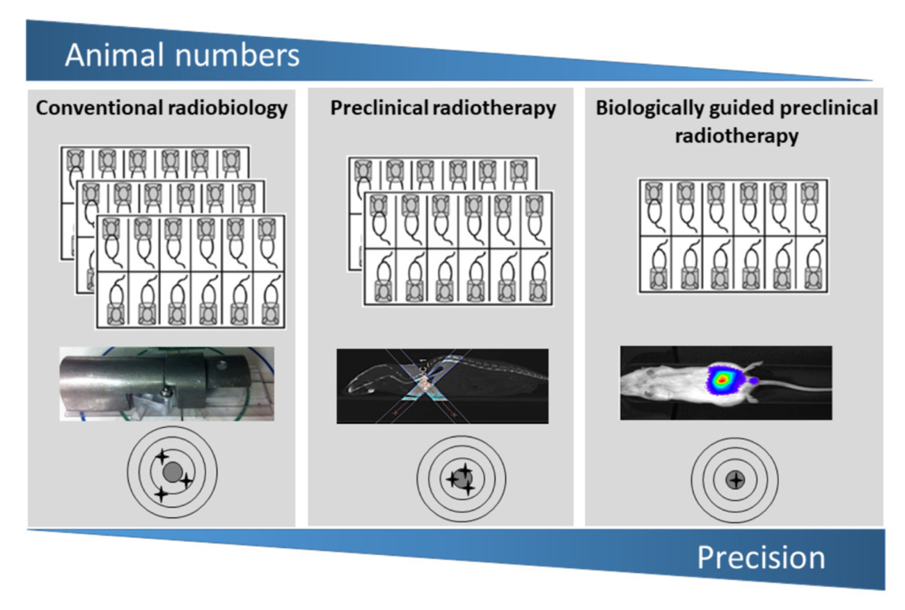
Mice are used to test the efficacy of new radiotherapy techniques. Clinical radiotherapy on humans uses specialised imaging to accurately identify a tumour within the surrounding tissue, allowing precise targeting of radiotherapy using small radiation fields. Due to the small size of rodents, this image guided radiotherapy is difficult to apply to preclinical radiotherapy studies, meaning mice may sustain additional soft tissue damage. Karl was awarded an NC3Rs PhD Studentship to reverse translate a clinically-validated solution to refine the use of mice in preclinical radiotherapy studies – an injectable marker that highlights tumour cells, called BioXmark. BioXmark is produced by Nanovi and was supplied for free for the PhD Studentship.
The 3Rs in synergy
Working with Karl, PhD Student Kathryn implemented and impacted all three ‘Rs’ in this project to overcome the challenges of using BioXmark to accurately target tumours during radiotherapy studies in small animals. During her PhD she was awarded best presentation at the Queen’s University Belfast Postgraduate Research Forum and published two first author papers [1,2]. Kathryn and Karl’s research highlights how the 3Rs integrate and influence each other in new approaches for better science.
Replace
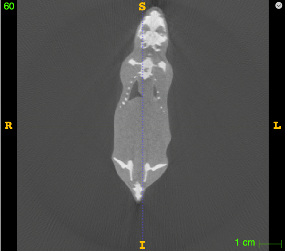
Before starting experiments with animals, the imaging machinery needs to be optimised. Instead of using animals to optimise the imaging parameters, Kathryn used ‘phantom’ measurements – this involves scanning a specially designed object to tune the performance of imaging devices. Mathematical simulations were then used to predict how tumour and normal tissues would respond to the radiotherapy.
Reduce
This computational data allowed Kathryn to calculate the number of animals required to determine an impact on the radiobiological response, meaning additional animals were not used unnecessarily. Preclinical studies that take up this technique will also require fewer animals as more accurate tumour targeting increases both the reproducibility and translatability of experiments.
Refine
Precisely targeting the tumour also minimises off-target soft tissue damage. Karl and Kathryn’s application of image guided radiotherapy devices to small animals is a major refinement that reduces the pain and suffering of animals in preclinical studies.
Promoting use through industry
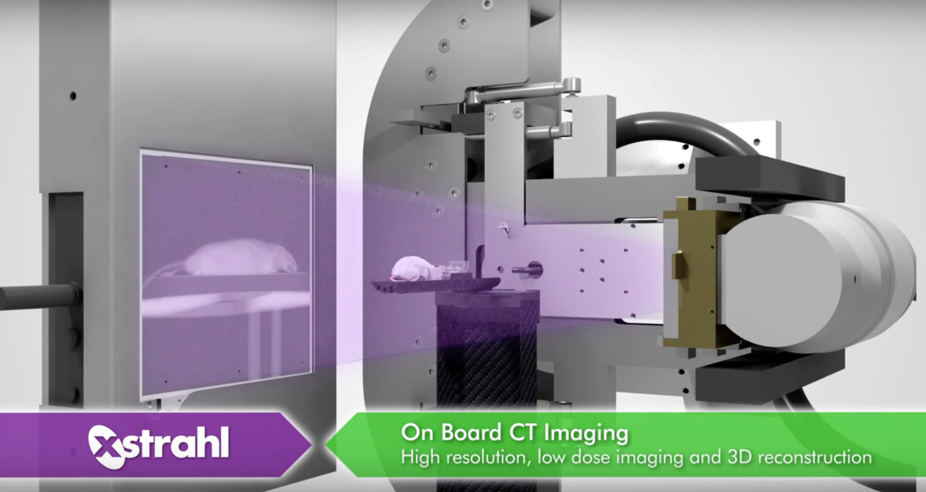
Karl has collaborated with radiotherapy systems provider XStrahl. Their small animal radiation research platform (SARRP) is specifically designed for preclinical research and allows scientists to adjust the radiation field to target areas as small as 1mm x 1mm. XStrahl hosted Karl for a webinar exploring how this project takes inspiration from clinical practice to offer innovative approaches for preclinical research.
As well as helping to facilitate the project by supplying BioXmark free of charge, Nanovi featured Kathryn’s publication in Cancers in a press release, raising awareness of the project among their business partners and end-users.
Supporting skills base for the future
After completing her PhD Kathryn was awarded an NC3Rs Training Fellowship and she is now developing computational models to extract enhanced data from existing imaging techniques, a technique known as radiomics. During her Fellowship, Kathryn has published four first author papers, sharing her research in a range of journals covering oncology and cancer, clinical and translational radiotherapy and research methods.
Kathryn was also awarded an Early Career Engagement Award to obtain further training, promote her 3Rs approach and in a collaborative effort with research groups in both the USA and Northern Ireland, Kathryn is also recreating an imaging phantom used for patient treatment for preclinical radiomics studies. These are specially designed objects used to evaluate and analyse the performance of imaging devices without needing patients, or animals, for this type of study. By using a standardised phantom of a clinical standard, Kathryn aims to better understand the similarities and differences between preclinical and clinical approaches.
Since receiving the Early Career Engagement Award, Kathryn has shared her work at three international conferences:
There’s been a lot of interest internationally in what I’m doing with the scanners to replace, reduce and refine animal use across all sorts of research areas. People I’ve met at these conferences are applying my approach to tissues and tumours in fields from fibrosis to longitudinal toxicity studies. It’s given me the chance to present to really different and mixed audiences, from benchtop researchers to clinicians to people working in mathematical modelling. A lot of contacts made and a lot of things added to the to-do list!
Dr Kathryn Brown, NC3Rs Training Fellow
References
- Brown K et al. (2020). Evaluation of a Novel Liquid Fiducial Marker, BioXmark®, for Small Animal Image-Guided Radiotherapy Applications. Cancers 12(5): 1276. doi: 10.3390/cancers12051276
- Brown K et al. (2022). A scoping review of small animal image-guided radiotherapy research: Advances, impact and future opportunities in translational radiobiology. Clinical and Translational Radation Oncology 34:112-19. doi: 10.1016/j.ctro.2022.04.004
Related projects