NC3Rs | 20 Years: Pioneering Better Science
Strategic grant

A tissue engineered construct to monitor mucosal immunity in asthma
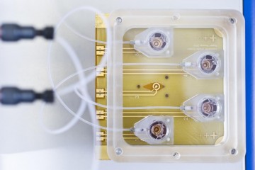
Monitor mucosal immunity in asthma

At a glance

Completed
Award date
January 2011 - March 2014
Grant amount
£499,713
Principal investigator
Professor Donna Davies
Institute
University of Southampton

R

  • Replacement

Application abstract

Asthma is an inflammatory disorder of the conducting airways which undergo distinct structural and functional changes leading to non-specific bronchial hyperresponsiveness (BHR) and airflow obstruction that fluctuates over time. It is amongst the commonest chronic conditions in Western countries affecting 1 in 7 children and 1 in 12 adults (equivalent to 5.1 million people in the UK) and is responsible each year for 1500 avoidable deaths, as well as 20 million lost working days. The annual UK health care cost is estimated to be £2.5 billion. A recent assessment of asthma across Europe (Brussels Declaration) has identified substantial unmet clinical need with those 10% of patients with severest disease accounting for ~50% of the health costs. It is of great concern that despite the wide use of ‘animal models of asthma' by the pharmaceutical industry, other than recombinant anti-IgE and anti-leukotrienes, both directed at targets identified many years ago, there have been no new therapeutic strategies for treating asthma since the introduction of ß2-agonists and corticosteroids in the 1960s. Many animal models have been used to study inflammation and alterations in airway function similar to those seen in human asthma. However, novel therapeutics developed using ‘animal models of asthma' have had disappointing outcomes when translated into the human disease. Alternative approaches for studying asthma mechanisms have employed in vitro or ex vivo models but even explanted tissue or tissue engineered constructs are limited by the absence of a circulation and the dynamic influx and efflux of leukocytes into the tissue which is a critical component of an inflammatory disease like asthma. Therefore, we aim to build on our previous NC3Rs-funded work to develop complex cellular models that utilize a microfluidic platform to enable delivery of inflammatory cells to a tissue engineered construct comprising endothelial and epithelial barriers with interstitial fibroblasts (FB). Our constructs will be integrated with an interactive monitoring system to assess barrier integrity, as well as pH, temperature, conductivity and O2 saturation of the buffer. The objectives of our proposed studies are: (a) To fabricate a microfluidic device combined with multi-parametric sampling and analysis to study the airway bronchial-vasculature barrier; (b) To develop a range of functional scaffolds that support differentiated epithelial and endothelial barriers for incorporation as a co-cultures into the microfluidic device; (c) To demonstrate proof-of-principle that the flow system can be used to assess cellular migration by measuring leukocyte migration through the epithelial-endothelial barrier; and (d) To validate the relationship between functional readouts in the tissue construct with responses observed in asthma; this will be achieved by measuring the effects of corticosteroids on barrier function, cytokine production and leukocyte influx. Thus, we anticipate that this system will allow us to integrate and validate corticosteroid-sensitive responses and to compare these with corticosteroid-refractory responses, where there is an unmet clinical need.

Impacts

Publications

  1. Blume C et al. (2017). Cellular crosstalk between airway epithelial and endothelial cells regulates barrier functions during exposure to double-stranded RNA. Immunity, inflammation and disease 5(1):45-56. doi: 10.1002/iid3.139
  2. Bucchieri F et al. (2017). Functional characterization of a novel 3D model of the epithelial-mesenchymal trophic unit. Experimental lung research 43(2):82-92. doi: 10.1080/01902148.2017.1303098
  3. Loxham M and Davies DE (2017). Phenotypic and genetic aspects of epithelial barrier function in asthmatic patients. Journal of Allergy and Clinical Immunology 139(6):1736-1751. doi: 10.1016/j.jaci.2017.04.005
  4. Blume C et al. (2015). Temporal Monitoring of Differentiated Human Airway Epithelial Cells Using Microfluidics. PloS One 10(10):e0139872. doi: 10.1016/j.jaci.2017.04.005
  5. Gordon S et al. (2015). Non-animal models of epithelial barriers (skin, intestine and lung) in research, industrial applications and regulatory toxicology. ALTEX 32(4):327-78. doi: 10.14573/altex.1510051
  6. Loxham M et al. (2014). Epithelial function and dysfunction in asthma. Clinical and Experimental Allergy 44(11):1299-313. doi: 10.1111/cea.12309
  7. Blume C et al. (2013). Barrier responses of human bronchial epithelial cells to grass pollen exposure. European Respiratory Journal 42(1):87-97. doi: 10.1183/09031936.00075612
  8. Blume C and Davies DE (2013). In vitro and ex vivo models of human asthma. European Journal of Pharmaceutics and Biopharmaceutics 84(2):394-400. doi: 10.1016/j.ejpb.2012.12.014
  9. Grainge CL and Davies DE (2013). Epithelial injury and repair in airways diseases. Chest 144(6):1906-1912. doi: 10.1378/chest.12-1944
  10. Leino MS et al. (2013). Barrier disrupting effects of alternaria alternata extract on bronchial epithelium from asthmatic donors. PloS One 8(8):e71278. doi: 10.1371/journal.pone.0071278