Malocclusion in mice
Information and resources on malocclusion in mice to improve welfare within a research setting.
Introduction
The information and resources on this page focus on malocclusion in mice and are intended to be used for training and education purposes to improve mouse welfare within a research setting. Although this resource focuses on mice, malocclusion also occurs in other rodents. Some of the information presented here may be generally applicable to other species, although species-specific guidance should also be consulted.
What is malocclusion?
Malocclusion is a dental disorder, common in laboratory mice and characterised by improper alignment of the teeth.
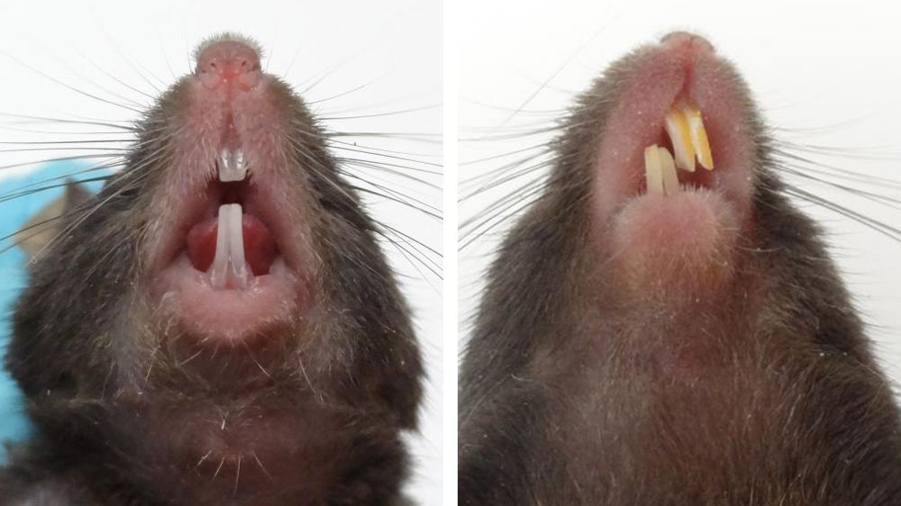
The teeth of rodents grow continually throughout their lifetimes and need to be worn down to prevent overgrowth. When the jaw of a healthy mouse is closed, the top and bottom incisors make contact with each other. When the teeth meet in this way, they wear down naturally as the mouse eats. If the top and bottom sets of teeth are not properly aligned, i.e. malocclusion is evident, the incisors become overgrown and this significantly impacts animal welfare.
The teeth of mice grow at a rate of 1 - 1.7 mm per week [1] therefore, if malocclusion is not addressed in its early stages, the animal is likely to experience suffering. Malocclusion can result in oral and facial abscesses, weight loss, malnutrition, dehydration and even death.
Risk factors associated with malocclusion
Malocclusion can result from misalignment of the jaw or uneven wear of the teeth. Malocclusion is often present from birth, but any mouse can develop malocclusion later in life.
Factors that increase the risk of malocclusion include:
- Gnawing very hard materials (e.g. cage lids, metal bars).
- Inappropriate feed (e.g. diet that is too soft).
- Blunt force trauma (e.g. through improper handling of mice or fighting with cage mates).
- Genetic background (e.g. mutations, pedigree).
- Certain models and procedures (e.g. oral tumour formation, irradiation).
Malocclusion cannot always be prevented, but steps can be taken to reduce its prevalence and the likelihood that it will develop into a serious condition:
- Regular welfare checks should include visual checks of the incisors.
- Gnawing of inappropriate materials should be addressed (e.g. through provision and assessment of environmental enrichment).
- Gnawing enrichment (e.g. nylon chews, wooden blocks) should be provided in sufficient quantities for all the mice within the enclosure.
- An appropriate solid diet should be provided (i.e. not just mash).
- Animals should always be handled carefully, confidently and securely using non-aversive methods. Mice habituated to non-aversive handling methods show increased willingness to be handled, which makes secure and confident handling an easier job.
- Extra care should be taken when handling pups.
- Mice should be monitored for signs of aggression and injury.
- Mice with malocclusion should not be used for breeding.
- Where practical and reasonable, the use of models, strains or procedures with an increased risk of malocclusion should be avoided.
- When it is known that the model, strain, or procedure puts the mouse at increased risk of malocclusion, the frequency of dental checks should be increased.
- All staff working with mice should be trained to spot signs of malocclusion and should know what steps to take if malocclusion is suspected or confirmed.
Checking for malocclusion
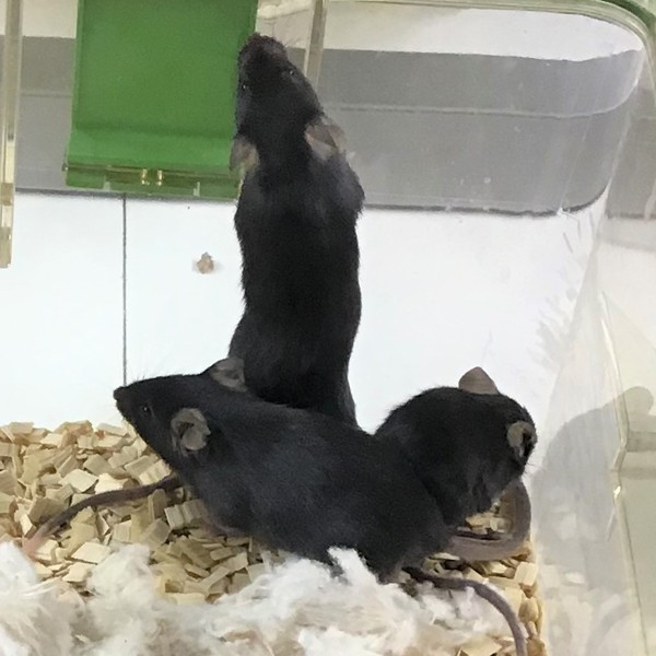
When making cage-side observations, look out for signs of general ill health such as:
- Smaller and/or thinner than cage mates.
- Smaller and/or thinner than littermates at weaning.
- Slower than cage mates; lethargic or not moving freely around the cage.
- Moving with an abnormal gait.
- Hunched posture.
- Abnormally shaped nose or face.
- Signs of pain evident in facial expressions (consult the mouse grimace scale).
Any of the above signs indicate that the mouse may be suffering from malocclusion; checking the teeth is necessary. To properly inspect the mouth and teeth you will need to scruff the mouse. Using non-aversive handling methods, as demonstrated in this video tutorial, reduces the impact of scruff restraint on mice.
Malocclusion is often evident around weaning age, or shortly after, when mice struggle to feed themselves. Increase vigilance for malocclusion around 2 - 5 weeks of age.
Malocclusion cannot always be prevented but spotting it in its early stages will prevent unnecessary animal suffering. Take time during welfare checks to confirm that teeth are:
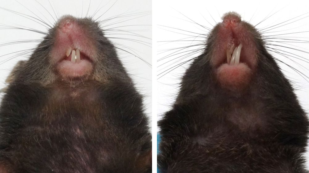
- Straight: the top and bottom incisors meet and do not grow at an angle.
- Intact: the teeth are not broken, and no uneven wear is evident.
- Normal length: there are no signs of overgrowth in either set of incisors, and the teeth do not make contact with any soft tissue.
If you are unsure if the teeth are healthy, ask for assistance in making this assessment.
Remember that the teeth of rodents grow quickly, and an early diagnosis can prevent the animal developing a serious health condition.
Taking action when malocclusion is evident
If malocclusion is evident, immediate action needs to be taken. It is important to know the procedure in your facility; however the process will usually follow the steps shown in the malocclusion decision tree, which can be printed for display in your facility. The text of the decision tree is outlined below.
Malocclusion is a serious welfare concern and warrants euthanasia. If keeping an animal is justified, a care plan will need to be put in place by the Named Veterinary Surgeon (NVS) and/or Named Animal Care and Welfare Officer (NACWO). This will vary depending on the circumstances but will usually require feeding the mouse on a soft or powdered diet and regular trimming of the teeth. It is important to use the correct tools for teeth trimming [2].
Malocclusion decision tree
This text is also available as a PDF diagram to download.
Physically assess the mouse. If it does not appear in good health*, or the known risk of malocclusion is high**, scruff to inspect the teeth.
- If teeth are healthy: investigate other potential causes of poor health following usual protocol.
- If malocclusion is evident (early or advanced stages): record health concern in notes/database.
- If animal is for stock/breeding: cull animal, record fate and details.
- If animal is for an experiment: contact researcher to discuss fate.
- If researcher requests to keep animal: contact named veterinary surgeon and/or animal welfare officer to discuss care plan.
- If researcher does not request to keep animal: cull animal, record fate and details.
*Signs of malocclusion include: smaller than cage/litter mates; underweight; lethargic; abnormally shaped nose/face; hunched posture; abnormal gait.
**Risk is increased for: pups around weaning age (2-5 weeks); irradiated animals; oral tumour models; genetically predisposed strains and pedigrees; animals without suitable gnawing enrichment.
Malocclusion training resources (including poster)
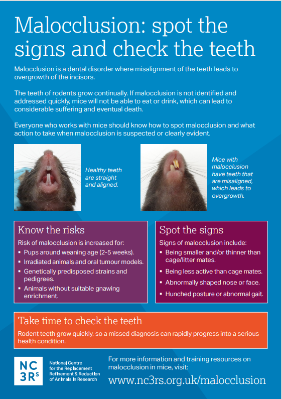
We have produced resources to help you spot the signs of malocclusion and take action, including a printable poster (please refer to the printing T&C below before printing).
Poster:
Available in German and English.
Decision tree and presentation slides:
Please note, the image below is included to demonstrate the content of the poster and should not be used for printing.
Poster printing terms and conditions
The proper use of the malocclusion poster requires the images to be clear and easily discernible. It therefore must be printed by a professional print service at the full A3 size. Further guidance is included in the cover page, which should not be removed from the PDF file.
Any requests to reproduce the poster, or to include it in any publications or training materials, should be directed to enquiries@nc3rs.org.uk. You should include how, why and where the poster will be used so that we can consider your case for approval. It is helpful to include any associated text, so we can see the context in which the poster will be put.
Copyright notice: This poster and its content are owned by the NC3Rs and its partners. It should not be adapted, and the content should not be sold or used to generate income.
References
- Coady JM, Toto PD, Santangelo MV (1967). Histology of the mouse incisor. Journal of Dental Research 46(2): 384-388. https://doi.org/10.1177/00220345670460021201
- Diagnosis | Severe prognathic malocclusion (2007). Lab Animal 36: 22–23. https://doi.org/10.0138/laban0107-22